PET/MRI Applications Update

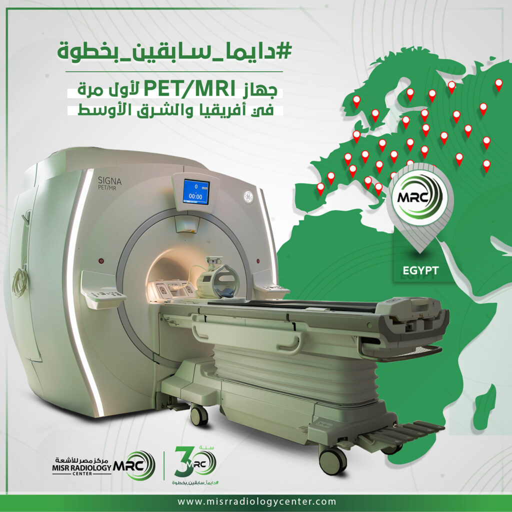
For the first time in Africa and Middle East, MRC is equipped with the latest technology PET/MRI for advanced diagnosis of various diseases.
PET/MRI combines the metabolic and molecular data of PET, with excellent anatomic details of MRI, using high end conventional MR protocols with all pertinent 3T sequences simultaneously in a one-stop-shop exam for accurate detection and characterization.
Following is some applications of the machine, with an overall update of where we stand:
1- Neurological applications of PET/MR:
1) Epilepsy: specially preoperatively and specially for children where PET/CT is not that favorable due to ionizing radiation.
- Also MR can provide structural data, together with MR perfusion and MR spectroscopy.
- Tracer used: FDG.
2) Brain tumors: specially evaluation of postoperative/posttherapy cases to differentiate viable tumor tissue from post treatment changes.
- The most appropriate tracer for this are choline tracers and also aminoacids tracers such as FET, F-DOPA tracers rather than FDG.
- Gallium-PMSA can be also used if the above are not present.
- The aminoacids tracers such as FET & F-DOPA are also useful for characterization and detection of low-grade gliomas unlike Gallium-PSMA which is applicable for high-grade tumors.
- Primary CNS lymphoma could be studied by FDG but Gallium-PSMA is better in this regard.
- Also in metastatic workup in known cases of primary cancer to evaluate the intracranial and intraspinal canal metastasis. This is usually done by FDG tracer.
- For meningioma in particular and also neuroendocrinal deposits, Gallium-DOTATATE is the preferred specific tracer and particularly when the patient has primary cancer and the aim is to differentiate meningioma from pachymeningeal metastatic deposit.
3) Neurodegenerative diseases including dementia cases:
- The applications for the above are widely increasing and we observe this not only for the evaluation of mild cognitive impairment/dementia like Alzheimer’s disease, frontotemporal dementia and lewy body dementia, yet because of the overlap of symptoms and cross correlations with other neurodegenerative diseases so evaluation of entities like traditional parkinsonism, parkinsonian plus syndromes for example multisystem atrophy and progressive supranuclear palsy and other forms of neurodegeneration such as corticobasal degeneration and posterior cortical atrophy are now widely evaluated by PET/MR specially that there could be combination of diseases for example Alzheimer with parkinsonism as well as CBD and LBD etc.
- For the above-mentioned causes of neurodegeneration, FDG is mainly used to assess the cases and especially that there is fingerprint like pattern for each case.
- Amyloid plaque tracer is used to rule out Alzheimer, if there is a negative amyloid scan, yet if there is positive scan, this can be with Alzheimer or other types of dementia so FDG is used to confirm whether it is Alzheimer or other type of dementia.
- For differentiation of traditional parkinsonism from parkinsonian plus cases, F-DOPA is used because there is high uptake in the basal ganglia in traditional parkinsonism while not in parkinsonian plus cases and this is important because the treatment is different.
4) Inflammatory lesions:
- Particularly autoimmune encephalitis and limbic encephalitis to differentiate from mesial temporal sclerosis because with encephalitis, the hippocampus is hypermetabolic, while with mesial temporal sclerosis, it is hypometabolic.
2- Body applications of PET/MR:
Generally speaking for children, it is always preferred to perform the study by PET/MR rather than PET/CT because of the ionizing radiation and specially if there is serial follow-up like in cases of lymphoma for example.
- The most important malignancies where PET/MR is used and even in some of them it is already in the guidelines for the year 2022:
- Prostate cancer (using PSMA).
- Rectal and anal cancers (using FDG).
- Urinary bladder cancer (using FDG).
- Gynecological malignancies (using FDG).
So from the above it is noted that pelvic cancers staging and follow-up is better done by PET/MR to combine the benefits of high end structural MR with diffusion and perfusion together with the PET data.
- Other cancers:
- Breast cancer staging.
- Liver cancer staging.
- Pancreatic cancer staging.
- Renal cancer staging.
The value is also to combine the high-end local staging as well as the whole-body staging.
- Generally speaking for multiple myeloma assessment and also similarly for melanoma assessment, PET/MR is preferred if present and applicable and this is because with PET/CT the lesions are detected but the specificity is less than MR that provides more specificity as in these cases there is overlap with inflammatory lesions and other lesions and MRI can solve these issues.
- For multiple myeloma in particular there are now better tracers than FDG like choline and methionine-based tracers and also carbon tracers but these need cyclotron presence in-house due to its short half-life.
- There is also use of ammonia tracers like for brain tumors and for cardiac applications, however, this also needs cyclotron in house due to a short half-life.
Note:
- Some centers due to less availability of PET/MR machines and insurance issues would do a local MRI study for the local staging for example for rectal cancer and then do a PET/CT for nodal and metastatic workup or staging, which is an inefficient way for diagnosis to all parties involved.
- When PET/CT is done or more preferred than PET/MR.
- Primary lung cancer.
- Upper GI cancers.
- Lymphoma initial staging and follow-up in adults (where the issue of ionizing ration is less important than in children).
- Contraindications for MR like pacemakers and aneurysmal coils…etc, or if the patient is obese and cannot fit in the 60cm bore of PET/MR or if he is uncomfortable or claustrophobic (anesthesia can be performed in these settings).
- No available time slots on PET/MR due to the much lesser availability of PET/MR scanners as compared to PET/CT scanners.
- Not enough professional MR readers that can do such hybrid or simultaneous reading of high-end MR combined with PET data and in this context, we noted that usually nuclear medicine experts are reading the PET/CT and the MR is read by radiologists whether neuroradiologist, body radiologist or cardiac radiologist…etc.
- If in post treatment follow-up, the patient had already performed high-end MRI for the local region and just needs follow-up for distant, nodal and metastatic deposits.
- However, in all these centers if there’s availability of doing the whole study in one stop shop by PET/MR, this is much preferred due to obvious reasons.
- Generally speaking in all centers visited globally, prostate cancer is reserved for PET/MR-Gallium PSMA, although some other tracers are now also tried.
4- Head and neck cancers:
Generally speaking and if available, it is preferred to perform PET studies for head and neck cancers by PET/MR because of its higher spatial resolution and ability to perform diffusion, perfusion and spectroscopy to the exam and last but not least to better assess for perineural spread of tumor.
5- Musculoskeletal applications of PET/MR:
These are not widely used except for malignancies and metastatic workup, yet people are enthusiastic about increasing their usage for inflammatory diseases specially in the acute stage and pinpointing the exact location and cause of painful conditions and to give you one example; not all back problems are due to disc herniations and not all joint pains are due to discal or ligamentous or tendineous injuries.
6- Cardiac applications of PET/MR:
As above-mentioned the main applications we observed are for ischemic heart disease using ammonia tracer to triage patients for better treatment but this can also be done by cardiac MRI and the other applications for entities like cardiac neoplasms, sarcoidosis, amyloidosis specially if there are equivocal cases after doing cardiac MRI and/or doing the study as PET/MR cardiac from the start combining all the techniques together.
Overall, the move towards adoption globally is strong and to give you practical example; in one of our site visits, the PET/MR list was as follows:
-
- Two cases for neurodegenerative disease/dementia.
- Two cases for prostate cancer.
- Two cases for breast cancer.
- One case of colon cancer.
- One case of liver cancer.
- Epilepsy cases and brain tumors cases are generally difficult for nuclear medicine experts because unlike neurodegeneration, the nuclear medicine people much need the presence of a professional neuroradiologist during the reading of these cases.
Finally, this is a summary of the current status of PET/MR applications in various body parts and I am ready to answer any questions for things not included in the above summary.
Much obliged,
Prof. Dr. Yasser Abd ELAzim
Professor of Radiology, Ain Shams University
Head of MRI & PET/MRI Units at Misr Radiology Center
If you have any questions, please feel free to reach out on medical_info@misrradiologycenter.com
July-2022
